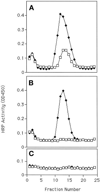
Figure 1.
Appearance of HRP chimeras in SLMVs after secretagogue stimulation. Pairs of 10-cm dishes were plated with PC12 cells transfected with ssHRPP-selectin (A and B) or TfnR-HRP (C) and incubated for 3 (A) or 7 d (B and C). One of each pair was then stimulated to secrete with carbamylcholine for 5 min, chased for 30 min, and then fractionated on a glycerol gradient (MATERIALS AND METHODS). The graph shows the amount of HRP activity in gradient fractions (MATERIALS AND METHODS). All points are the mean of triplicate assays for that fraction. Error bars indicate the SD for the intraexperimental variation. Note that in this and most subsequent figures, the variability of the HRP assay is so low that no error bars can be seen at the scale of reproduction used here. Interexperimental variation is detailed in the text. The effectiveness of stimulation was monitored in all cases by preloading with 3H-dopamine and counting media and cell samples. Fraction 1 is at the top of all gradients. □, Unstimulated; ♦, stimulated.

