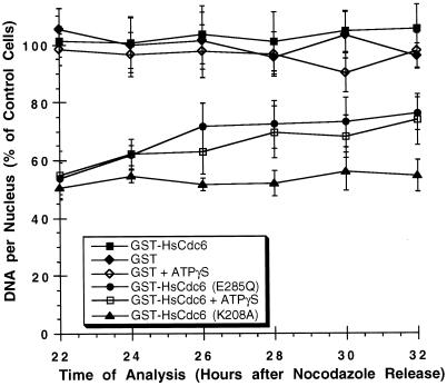
Figure 9.
Walker A mutants of HsCdc6 arrest cells before S phase, and Walker B mutants prevent completion of S phase. HeLa-S3 cells that had been arrested in G2/M with nocodazole for 16 h were released into nocodazole-free medium for 6–8 h. The following proteins at the indicated concentrations were microinjected into the nuclei of the cells: GST (500 ng/μl; filled diamonds), GST-HsCdc6 (61 ng/μl; filled squares), GST-HsCdc6 (K208A) (20 ng/μl; filled triangles), GST-HsCdc6 (E285Q) (30 ng/μl; filled circles), GST-HsCdc6 (109 ng/μl) preincubated with a twofold molar excess of ATPγS (open squares), and GST (500 ng/μl) preincubated with the same concentration of ATPγS used for GST-HsCdc6 (open diamonds). At 12 h after the release, the medium was again supplemented with nocodazole to prevent progression through mitosis. At the indicated times, the cells were stained with anti-GST polyclonal antibody and FITC-conjugated goat anti-rabbit secondary antibody and Hoechst 33258 fluorochrome. Nuclear DNA content was measured by fluorescence microscopy. Nuclear DNA content of injected cells is expressed as a percentage of the nuclear DNA content of uninjected cells in the same field of vision, which was set to 100%. For each time point, the average value obtained from at least 10 cells is shown. The SD of the mean is indicated by error bars.

