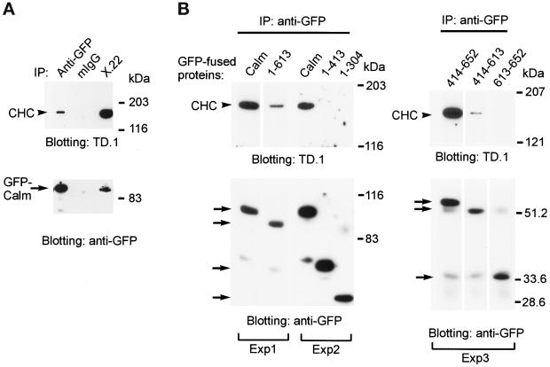
Figure 10.
Coimmunoprecipitation of CHC with CALM and its fragments. (A) The lysates of COS-1 cells transfected with GFP–CALM were incubated with anti-GFP, X.22, or nonspecific mouse IgG (mIgG), and the immunoprecipitates were resolved by SDS-PAGE. CHC and GFP–CALM were detected by blotting with TD.1 or anti-GFP, respectively. (B) GFP–CALM, GFP–414–652, GFP–1–413, GFP–1–304, GFP–1–613, GFP–414–613, and GFP–613–652 fragments (see Figure 2) were immunoprecipitated with anti-GFP from the TX100 lysates of transiently transfected COS-1 cells. The immunoprecipitates were analyzed by SDS-PAGE, and the presence of CHC and GFP fusion proteins in the immunoprecipitates was detected by blotting with antibodies TD.1 or anti-GFP, respectively. Arrows indicate the positions of the GFP fusion proteins, whereas the arrowheads point to the position of CHC. The lanes in each panel (labeled Exp1, Exp2, and Exp3) are from the same experiment. The data in each panel are representative of several independent experiments.

