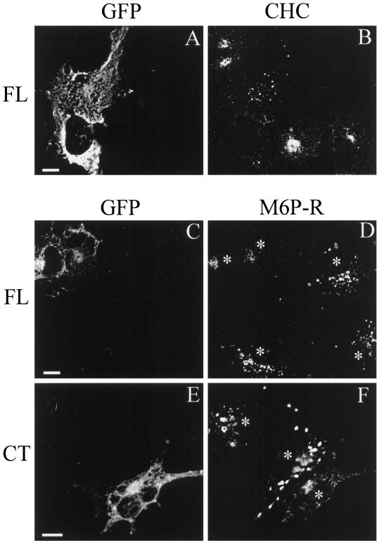
Figure 9.
The effect of GFP–CALM expression on the localization of clathrin and M6P receptor. COS-1 cells expressing GFP–CALM (FL; A–D) or GFP–414–652 (CT; E and F) grown on coverslips were fixed and stained with mouse monoclonal anti-clathrin X.22 (A and B) and rabbit polyclonal anti-M6P receptor (C–F) followed by the secondary goat IgGs specific to mouse or rabbit IgG, respectively, labeled with Texas Red. The serial optical sections were acquired through the Texas Red (red) and GFP (green) channels and deconvoluted as described in MATERIALS AND METHODS. Images represent the merged images of two serial optical sections (total thickness of 0.4 μm) from the middle of the cell, where the most intense signal for CHC or M6P receptor (M6P-R) in the perinuclear area was observed. Asterisks show the position of cell nuclei. Bars: A and B, C and D, and E and F, 10 μm.

