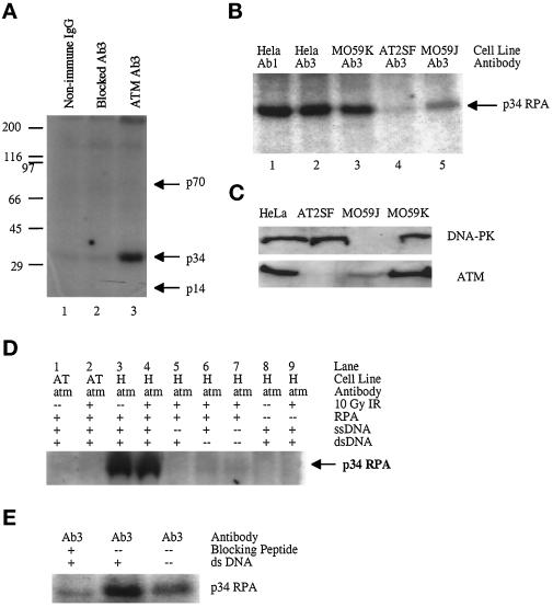
Figure 6.
Phosphorylation of p34 RPA by ATM-associated protein kinase. All kinase reactions were performed with immunoprecipitates from equal amounts of cellular protein. (A) ATM immunoprecipitated from HeLa cells with nonimmune rabbit IgG (Nonimmune IgG, lane 1), peptide-blocked Ab3 (Blocked, lane 2), or Ab3 (ATM Ab3, lane 3) and incubated with [γ-32P]ATP, ssDNA, dsDNA, and purified RPA. (B) ATM protein kinase activity immunoprecipitated from HeLa (lanes 1 and 2), MO59K (lane 3), AT2SF (lane 4), and MO59J (lane 5) cells. (C) Lysate (50 μg) from HeLa (lane 1), AT2SF (lane 2), MO59J (lane 3), and MO59K (lane 4) cells probed with antibodies to DNA-PK (top) or ATM Ab3 (bottom). (D) ATM-associated protein kinase activity in AT2SF cells (AT) (lanes 1 and 2) or in HeLa cells (H) (lanes 3–9) with purified RPA used as a substrate (lanes 1–7). (E) Kinase reactions performed on immunoprecipitates from MO59J cells with blocked Ab3 (lane 1) and Ab3 (lanes 2 and 3) in the presence (lanes 1 and 2) or absence (lane 3) of dsDNA.

