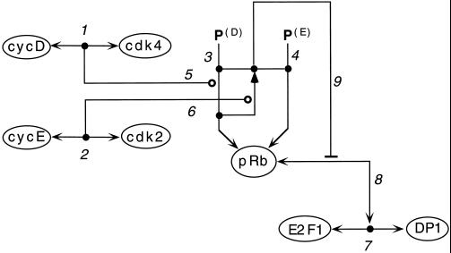
Figure 5.
Phosphorylation control of pRb: illustration of the use of conjunction symbols to denote protein modification combinations. (1) Cyclin D binds Cdk4; the filled circle (node) on the line represents the CycD:Cdk4 complex itself. (2) Similarly for Cyclin E and Cdk2. (3) The single-arrowed line linking P(D) to pRb represents phosphorylation of pRb at sites kinased by CycD:Cdk4; a node on this line represents pRb-P(D) [the P(D)-phosphorylated form of pRb]. (4) Similarly for pRb phosphorylated at sites, P(E), kinased by CycE:Cdk2. (5) CycD:Cdk4 phosphorylates pRb at sites P(D). (6) CycE:Cdk2 acts on pRb-P(D), generating fully phosphorylated pRb; the line with the filled arrowhead indicates stoichiometric conversion of pRb-P(D) to the fully phosphorylated form, which is represented by the node on the nonarrowed line connecting the pRb-P(E) and pRb-P(E) nodes. (7) E2F1 binds DP1. (8) pRb binds to E2F1:DP1. (9) Fully phosphorylated pRb can-not bind to E2F1:DP1. Thus dissociation of pRb from E2F1:DP1 requires hyperphosphorylation by Cyclin E:Cdk2, which in turn requires previous phosphorylation by Cyclin D:Cdk4/6 (Zarakowska and Mittnacht, 1997; Lundberg and Weinberg, 1998).

