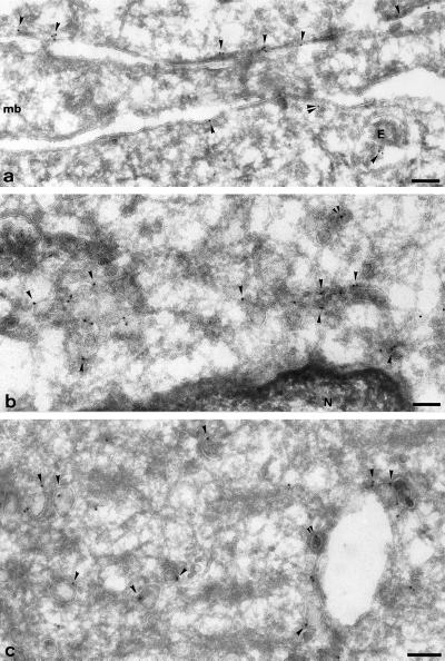
Figure 12.
Ultrastructural localization of VAMP5. Ultrathin frozen sections of differentiated C2C12 cells were labeled with anti-VAMP5 antibodies followed by swine anti-rabbit antibodies and then 10 nm protein A-gold. Panel a shows plasma membrane regions of three closely apposed cells; a region of myoblast (mb) is unlabeled, whereas the neighboring myotubes (above and below) show significant labeling close to the plasma membrane (arrowheads). Note the labeling on a small vesicle (double arrowheads) and on a multivesicular endosome (E). Panel b shows the juxtanuclear area of a myotube; labeling is concentrated on uncoated vesicular and tubular profiles. The ER surrounding the nucleus (N) is unlabeled. Panel c shows characteristic labeling of large uncoated vesicles (arrowheads), some of which are associated with putative endosomal structure. The double arrowheads in b and c indicate a labeled clathrin coated buds. Bars, 100 nm.

