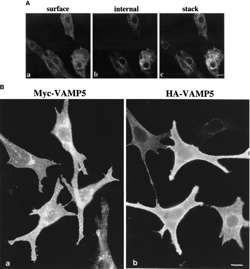
Figure 7.
(A) Labeling of VAMP5 in few myoblasts in differentiated C2C12 cultures by indirect immunofluorescence microscopy. Cells were fixed, permeabilized, and incubated with affinity-purified rabbit antibodies against VAMP5 followed by FITC-conjugated anti-rabbit IgG. The cells were mounted, and images captured with the Bio-Rad MRC1024 confocal system. Although the majority of the unfused myoblasts did not exhibit any specific labeling, a small fraction of unfused myoblasts had strong labeling on the cell surface (A, a) and intracellular vesicular structures (A, b) when viewed at the cell surface and internal focal planes, respectively. Shown in c (A) is the combined image of 0.3-μm optical sections, and it is again obvious that VAMP5 is associated with the plasma membrane and intracellular vesicular structures. (B) Epitope-tagged versions of VAMP5 are targeted to the cell surface. Pooled transfectants of C2C12 cells stably transfected with expression vector expressing myc-tagged VAMP5 (a) or HA-tagged VAMP5 (b) were fixed and labeled with monoclonal antibody against myc or HA followed by FITC-conjugated anti-mouse IgG. Cells were viewed and photographed. Bars, 10 μm.

