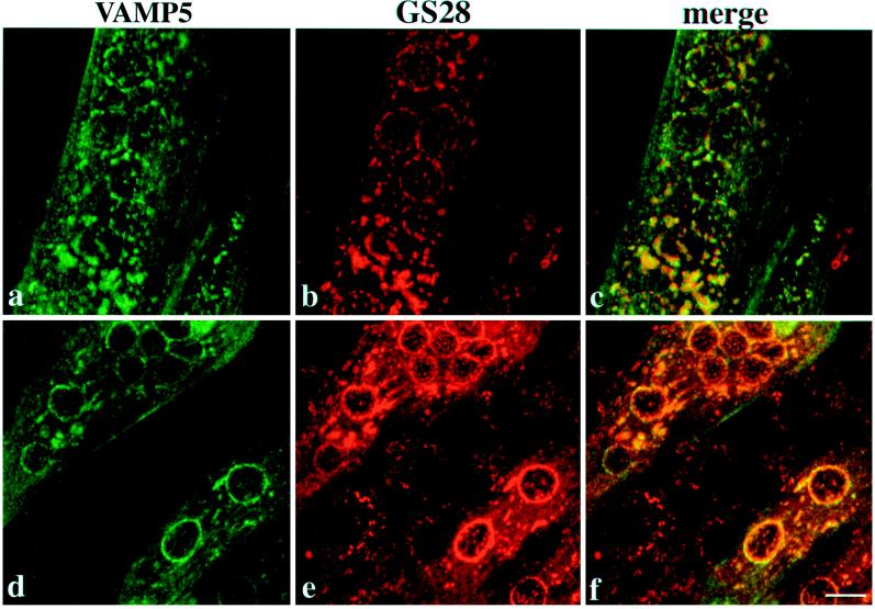
Figure 9.
Differentiated C2C12 cells were incubated with 10 μg/ml nocodazole for 60 min and then analyzed by indirect immunofluorescence microscopy to detect VAMP5 and GS28. The top and bottom panels represent views from different myotubes. Note that in the bottom panel, unfused myoblasts are labeled by anti-GS28 antibody (e and f) but are negative for VAMP5 labeling (d and f). Although the Golgi apparatus marked by GS28 in unfused myoblasts is clearly fragmented, the Golgi apparatus marked by GS28 in myotubes is less fragmented. Under this condition, the Golgi-like labeling of VAMP5 is significantly segregated from that of GS28. Bar, 10 μm.

