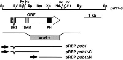
Figure 1.
Restriction map of the pob1 locus. Restriction sites on the insert of pWT4–3 are shown on the top. The extent and direction of the pob1 ORF, encoding 871 amino acids, are indicated by the arrow. The hatched box on the arrow indicates an SH3 domain, the shaded box indicates a SAM domain, and the filled box indicates a PH domain. The structure of a linear fragment carrying ura4+, which was used for disruption of the pob1 gene in vivo, is illustrated under the arrow. Inserts of three groups of subclones carrying the whole or a part of the pob1 ORF, collectively denoted either pREPpob1, pREPpob1ΔC, or pREPpob1ΔN, are also indicated. Restriction sites: Bg, BglII; Bm, BamHI; EI, EcoRI; EV, EcoRV; Hc, HincII; Pv, PvuII; Nd, NdeI; Sa, SacI; Sc, ScaI; Sp, SpeI; and Xb, XbaI.

