Figure 5.
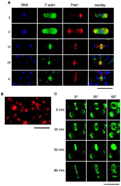
Localization of Pob1p. (A) Comparison of intracellular localization of Pob1p and F-actin. Haploid pob1Δ cells expressing HA-Pob1p were fixed in growth phase and stained with anti-HA to detect Pob1p, BODIPY-phallacidin to detect F-actin, and Hoechst 33342 to detect DNA. The panels show from left to right: localization of nuclei (DNA) displayed in blue, localization of F-actin displayed in green, localization of Pob1p displayed in red, and overlays of these three images. (B) Detection of Pob1p-3HA expressed from a single-copy chromosomal gene. Cells of JW100 were grown asynchronously in liquid YE medium at 30°C, fixed and stained with anti-HA. (C) Chronologically chased images of growing JX1001 cells. These cells were allowed to express an appropriate level of GFP-tagged Pob1p by adding a limited amount of thiamine, so that they would assume morphology close to the wild-type. GFP fluorescence was digitized and recorded by confocal microscopy. The original view (0°) and synthesized images after rotation of either 30° and 60° are shown. Bar, 10 μm.

