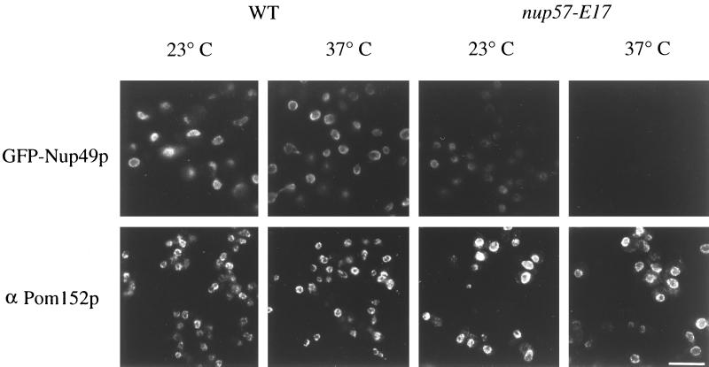
Figure 6.
Localization of GFP-Nup49p at the NPC is decreased in nup57-E17 cells, whereas Pom152p localization is not perturbed. Within each row, images were photographed and printed for identical times for direct comparison of fluorescence intensities. Top row, direct fluorescence microscopy was performed on wt and nup57-E17 cells expressing GFP-Nup49p (SWY809 and SWY1586, respectively), grown in SD lacking tryptophan. At 23°C, the nup57-E17 strain has less GFP fluorescence signal localized at the NE than wt cells. After 4 h of growth at 37°C, the nup57-E17 cells lack any detectable GFP-Nup49p staining. Bottom row, Indirect immunofluorescence microscopy was performed with monoclonal anti-Pom152p antibodies (mAb 118C3) on wt (SWY519) and nup57-E17 (SWY1587) strains grown in YPD. The anti-Pom152p staining is localized at the NE/NPC, and the intensity level is not notably altered in nup57-E17 cells at either 23 or 37°C compared with wt. Bar, 10 μm.

