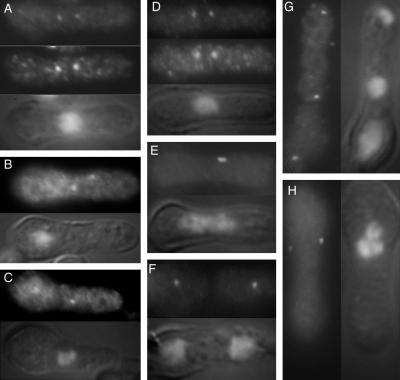
Figure 9.
Plo1 associates with the SPB of stf1/cut12 mutants and Stf1/Cut12 associates with the SPB of cells lacking Plo1. (A–C) Germinating spores from a homozygous ura4.d18 diploid strain in which one copy of the stf1/cut12+ gene had been replaced with the ura4+ gene were inoculated into media lacking uracil and incubated at 30°C for 20 h before processing for immunofluorescence microscopy. The top panels show anti-Plo1 staining, whereas the middle panel shows the result of simultaneous staining with a sheep anti-Sad1 antibody. The bottom panel in each image is a combined DAPI/Nomarski image of the cell in the other panels. The cells in B and C were stained only with Plo1 antibodies. Plo1 localizes to each SPB in all cases, regardless of their lack of Stf1/Cut12 and the inherent inability to execute mitosis. (D–H) Germinating spores from a homozygous ura4.d18 diploid strain in which one copy of the plo1+ gene had been replaced with the ura4+ gene were inoculated into media lacking uracil and incubated at 30°C for 14 h before processing for immunofluorescence microscopy. The top panels in D–F and the left-hand panels in G and H show anti-Stf1/Cut12 staining, whereas the bottom panel in D–F and the right-hand panels in G and H show DAPI DIC images. The middle panel in D shows the result of simultaneous staining with a sheep anti-Sad1 antibody. The cells in E–H were stained only with Stf1/Cut12 antibodies. Stf1/Cut12 localizes to each SPB in all cases, regardless of their lack of Plo1.

