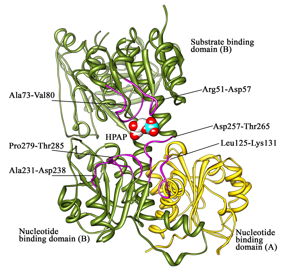
Figure 1. M.tb PGDH active site.
The figure shows the loops bordering the active site of M. tb PGDH in chain B in purple. The active site is formed by both chains. The nucleotide and substrate binding domain of chain B are shown in dark green. The intervening and regulatory domains are not shown for clarity. The nucleotide binding domain of chain A is in gold. The substrate, HPAP, is shown in space filling representation bound at the active site (carbon atoms are white, oxygen atoms are red, and phosphorus is cyan).

