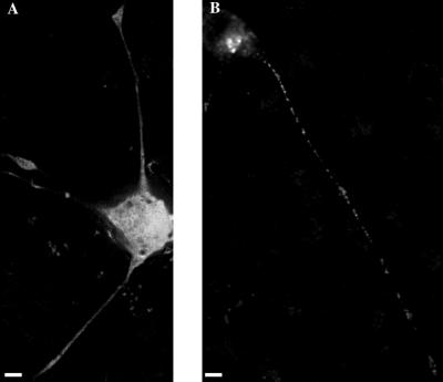
Figure 5.
Deblurred images of fixed (A) tPA/GFP(SIG−) and (B) GFP(SIG+)-expressing PC12 cells. One significant difference in distribution between tPA/GFP and GFP(SIG+) is that the staining in growth cones and neurites is often so faint in GFP(SIG+)-expressing cells that these structures are almost invisible when viewed in fluorescence mode. The GFP(SIG+) labeling in the neurite and growth cone shown here is somewhat more intense than is typical, permitting these structures to be seen in the image. Differences in growth cone brightness exhibited by tPA/GFP and GFP(SIG+) are not due to a lower level of expression in GFP(SIG+)-expressing cells. In fact, GFP(SIG+)-expressing cells on average are brighter than tPA/GFP-expressing cells. Bar, 4 μm.

