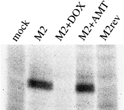
Figure 1.
Adenoviral expression of Rostock M2 in polarized MDCK cells. MDCK T23 cells grown on filter inserts (12 mm diameter) were mock infected (mock) or infected for 60 min at 37°C with 2 μl AV-M2 (M2) or AV-M2rev (M2rev). DOX (20 ng/ml) or AMT (5 μM) was added to the medium immediately after removal of the virus. The following day, cells were starved for 30 min and pulse labeled for 2 h with [35S]methionine in the continued presence of either DOX (M2+DOX) or AMT (M2+AMT). Filters were solubilized, the lysates were centrifuged briefly to remove debris, and M2 was immunoprecipitated. Samples were electrophoresed on a 12% SDS-polyacrylamide gel, and the dried gel was exposed to x-ray film.

