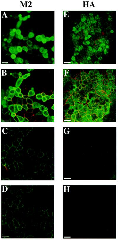
Figure 2.
Localization of M2 and HA in polarized MDCK cells using indirect immunofluorescence. Polarized MDCK T23 cells were infected with AV-M2 (A–D) or AV-HA (E–H) as described in MATERIALS AND METHODS. The following day, cells were fixed, permeabilized, and double labeled using antibodies against ZO-1, a component of tight junctions (A–H, red), and either M2 (A–D, green) or HA antibody (E–H, green). Individual confocal sections taken from the apical surface (A and E), the level of the tight junction (B and F), the lateral surface (C and G), and the basal surface (D and H) are shown. Adenoviral infection does not disrupt the polarized morphology of the cells, because tight junction staining is normal, and HA is localized almost exclusively to the apical surface. By contrast, M2 is concentrated at the apical domain, but lateral and basal staining is also evident. Bars, 10 μm.

