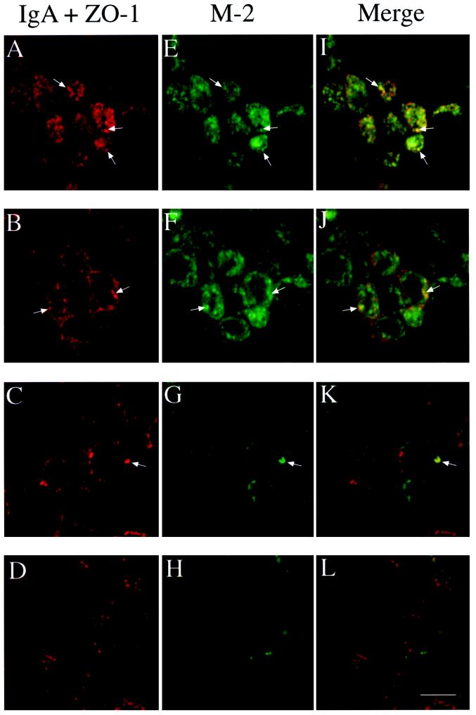
Figure 3.
Intracellular M2 partially colocalizes with internalized IgA in AREs. Filter-grown T23 cells infected with AV-M2 were allowed to internalize IgA from the basolateral side for 10 min at 37°C and then washed extensively and chased for 3 min to accumulate internalized IgA in the ARE. Cells were then chilled, trypsin-treated, fixed, permeabilized, and processed for indirect immunofluorescence. Individual confocal sections taken from the apical surface (A, E, and I), the level of the tight junction (B, F, and J), the lateral surface (C, G, and K), and the basal surface (D, H, and L) are shown. M2 (E–H) was localized using FITC-conjugated secondary antibody, and IgA and ZO-1 (A–D) were detected using Cy-5–conjugated secondary antibody. (I–L) Merged images. Arrows point to examples of structures in which M2 and IgA colocalize. Bar, 10 μm.

