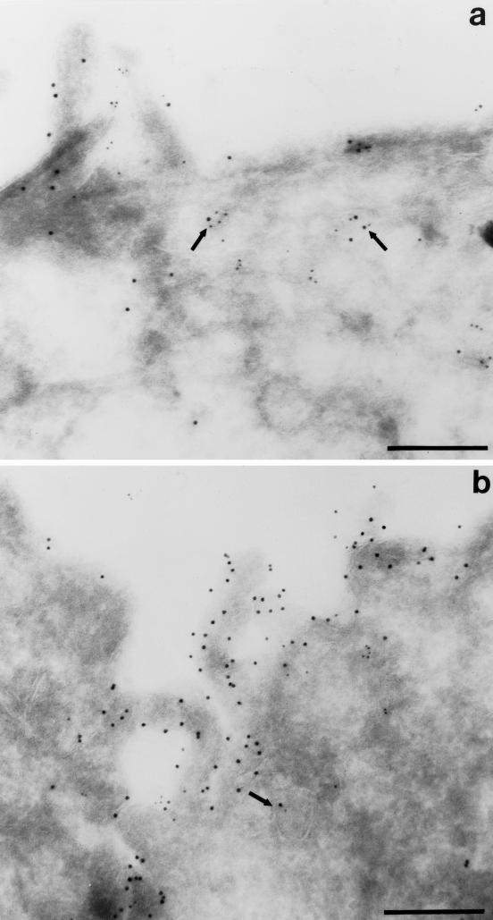
Figure 4.
Colocalization of M2 and internalized IgA by indirect immunoelectron microscopy. Filter-grown T23 cells infected with AV-M2 were allowed to internalize IgA from the basolateral side for 10 min at 37°C and then washed extensively and chased for 3 min. Cells were then processed for double-label immunoelectron microscopy. M2 was detected using 10 nm gold, and IgA was localized using 5 nm gold. Subapical vesicles containing both M2 and IgA are marked with arrows. Bar, 0.25 μm.

