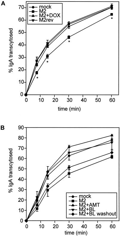
Figure 5.
M2 slows the rate of basolateral-to-apical transcytosis of IgA. (A) Filter-grown MDCK T23 cells were mock infected or infected with AV-M2 or AV-M2rev and then treated with 2 mM butyrate (to induce pIgR synthesis) in the presence or absence of 20 ng/ml DOX. The next day, cells were incubated with basolaterally added [125I]IgA at 37°C for 10 min and then washed extensively to remove free [125I]IgA. After warming, the appearance of [125I]IgA in the apical medium was monitored over a 1-h period. The mean ± SD from triplicate samples for each condition is shown. *, p ≤ 0.005 compared with mock-infected controls. Similar results were observed in at least six experiments for each condition. (B) Filter-grown MDCK T23 cells were mock infected or infected with AV-M2, and the reversible M2 inhibitor BL-1743 (5 μM) was added to the indicated filters immediately after infection. The next day, filters were incubated for 30 min at 37°C in MEM/BSA before [125I]IgA uptake. During this period, BL-1743 was washed out from some of the pretreated filters (M2+BL washout, diamonds), and AMT (5 μM) was added to some of the previously untreated filters (M2+AMT, triangles). The cells were then incubated with basolaterally added [125I]IgA at 37°C for 10 min, and IgA transcytosis was monitored in the continued presence of BL-1743 (M2+BL, upside-down triangles) or AMT. The average ± range from duplicate samples is shown. Similar results were obtained in three experiments.

