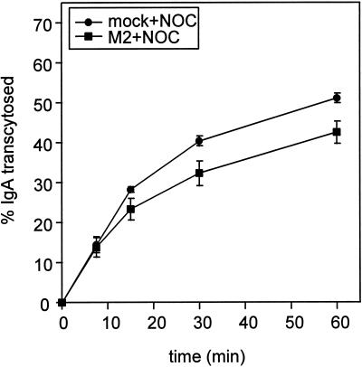
Figure 6.
M2 blocks IgA transcytosis after the microtubule-dependent step. Filter-grown mock-infected or AV-M2-infected MDCK T23 cells were incubated with basolaterally added [125I]IgA for 10 min and then rapidly chilled and washed extensively. The cells were then warmed to 37°C for 3 min to chase [125I]IgA into the ARE, rapidly chilled, and incubated in NOC-containing medium on ice for 60 min. After warming in the continued presence of NOC, release of transcytosed [125I]IgA into the apical medium was monitored as described above. The mean ± SD from triplicate samples is shown. Similar results were obtained in four experiments.

