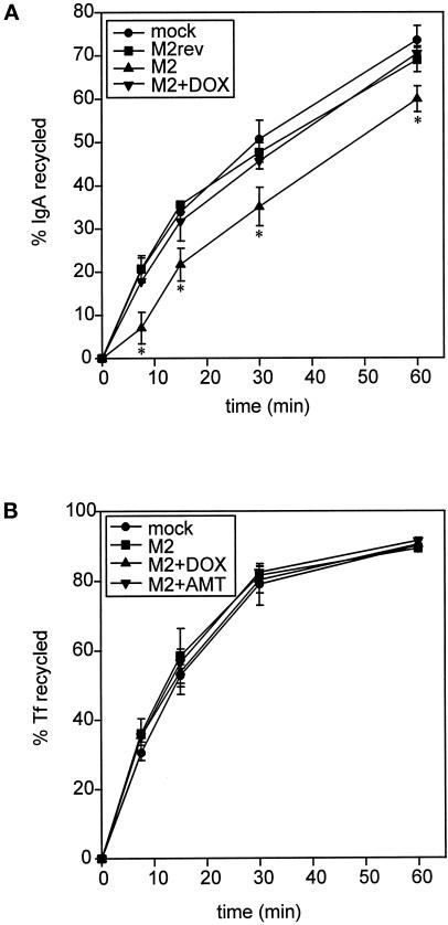
Figure 7.
M2 expression affects apical but not basolateral recycling in polarized MDCK T23 cells. (A) MDCK T23 cells were mock infected or infected with AV-M2, and the indicated samples were treated with DOX overnight. Cells were incubated with apically added [125I]IgA for 30 min and washed extensively at 0°C, and reappearance of endocytosed pIgA into the apical medium was monitored at 37°C. The average ± range from duplicate samples is plotted. *, p < 0.05 compared with mock-infected controls (data from 4 experiments compared using paired t test). (B) MDCK T23 cells infected as above were incubated with basolaterally added iron-loaded [125I]Tf for 10 min and washed extensively, and basolateral recycling of the preendocytosed [125I]Tf was monitored as described in MATERIALS AND METHODS. The mean ± SD from triplicate samples is shown. Similar results were obtained in four experiments.

