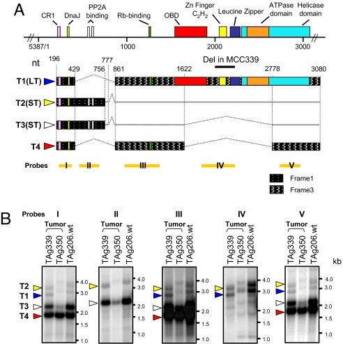
Fig. 1.
Transcript mapping of the multiply spliced MCV T antigen locus. (A) RACE analysis on MCC tumors shows four major MCV T antigen transcripts: T1 (LT, blue arrow), T2 (ST, yellow), T3 (ST, white), and T4 (17kT analog, red). Straight and dotted lines indicate noncoding and spliced regions, respectively. All four transcripts encode CR1 (pink, LXXLL) and DnaJ (light green, HPDKGG) domains. ST protein contains two PP2A binding motifs (white, CXCXXC). LT protein contains Rb binding (green, LXCXE), origin binding (red), zinc finger (yellow), leucine zipper (blue), and helicase (cyan)/ATPase (orange) motifs. The 17kT analog protein also encodes the Rb-binding motif. (B) Total RNA extracted from MCV T antigen-transfected 293 cells was Northern-hybridized to probes corresponding to mapped exons and introns (I–V in A). Each colored triangle indicates the corresponding transcript shown in A. An MCV339 deletion shortens TAg 339 T1 and T2 transcripts but is spliced out of T3 and T4 transcripts, which are similar in length to TAg350 and TAg206 T3 and T4 transcripts.

