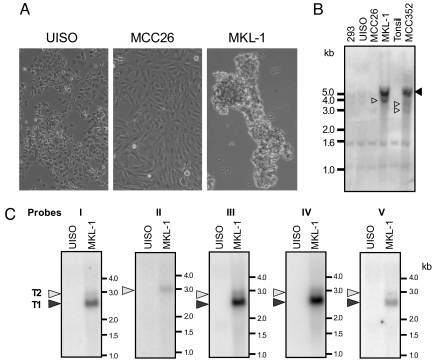
Fig. 3.
Identification of MCC line MKL-1 with monoclonal MCV-integration. (A) Uninfected variant (UISO, MCC26) MCC cells have a flattened, adherent morphology, whereas MCV-positive classic (MKL-1) MCC cells are rounded and loosely adherent. Phase-contrast images are at ×100 magnification. (B) Southern blot for MCV genomic DNA in cell lines (MCC352 tumor as positive control). Closed triangle (5.1 kb) represents viral genome in head-to-tail concatemers, and open triangles are viral-host fusion fragments. (C) Northern hybridization of UISO and MKL-1 total RNA with probes shown in Fig. 1A demonstrates expression of T1 (LT) and T2 (ST) transcripts in MLK-1 cells.

