Fig. 4.
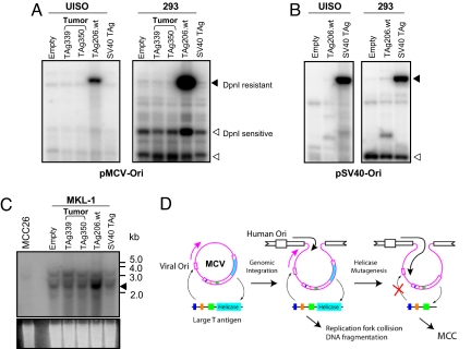
Initiation of MCV replication by WT but not tumor-derived LT. (A) T antigen expression vectors (TAg339, TAg350, and TAg206.wt) or pcDNA vector (Empty) were cotransfected with MCV replication origin plasmid (pMCV-Ori) in UISO and 293 cells, and replication was detected by Southern blotting. The positions of replicated DNA (DpnI-resistant) and unreplicated DNA (DpnI-sensitive) are indicated by solid and open arrows, respectively. (B) SV40 LT expression vector and SV40 origin plasmid were used as a positive control for the replication assay. Neither MCV nor SV40 LT initiates replication from each others' viral origin. (C) Southern blot of MKL-1 cells for MCV VP1 gene shows WT 206LT, but not tumor-derived 339LT or 350LT, induces endogenous integrated MCV DNA replication (arrow) after transfection. MCC26 was used as negative control for this replication assay. EtBr-stained agarose gel is shown as a loading control. (D) MCV integration into host chromosomes can be expected to lead to autonomous viral origin DNA replication when WT T antigen is expressed. Newly replicated virus DNA strands may collide with cellular replication forks unless secondary mutations eliminate viral LT antigen helicase activity.

