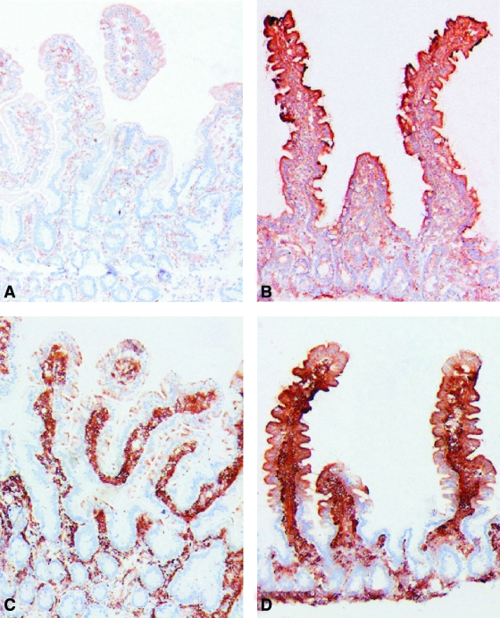
FIG. 3.
Immunoperoxidase stainings for HLA-DR (A and B) and HLA-DP (C and D) in jejunal biopsy specimens from a healthy control (A and C) and from a type 1 diabetic patient with normal jejunal mucosa (B and D). Intensive, positive HLA-DR staining is seen throughout the epithelial cells of the villi and also in many crypt cells in the type 2 diabetic specimen (B), whereas the control specimen shows only faint HLA-DR staining in the apical parts of the epithelial cells at the tip of the villi (A). The biopsy specimen from a control patient treated with HLA-DP antibody shows scattered positive granules in the apical parts of the epithelial cells at the tip of the villi (C), whereas strong positive staining is seen throughout the villous epithelial cells in the specimen from a type 1 diabetic patient (D). No positive staining is seen with either HLA-DR or HLA-DP in crypt cells in the control specimen (A and C). AEC-hematoxylin stain, original magnification ×50. (Please see http://dx.doi.org/10.2337/db08-0331 for a high-quality digital representation of this figure.)

