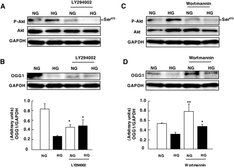
FIG. 4.
Role of PI 3-kinase activation by high glucose (HG) on OGG1 expression in MCT cells. A and C: Representative immunoblot shows a decrease in phospho-Akt (p-Akt) expression in cells preincubated with 50 μmol/l LY294002 and 100 nmol/l Wortmannin, respectively, before exposure to high glucose for 60 min. B and D: Representative immunoblot shows an increase in OGG1 expression in cells preincubated with 50 μmol/l LY294002 and 100 nmol/l Wortmannin before exposure to high glucose for 60 min. Histograms in the bottom panel represent means ± SE of three independent experiments. Significant difference from nontreated cells is indicated by *P < 0.05 and **P < 0.01. NG, normal glucose.

