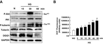
FIG. 8.
Effect of high glucose (HG) concentration on Akt/PKB and tuberin phosphorylation and OGG1 expression in primary cultures of RPTE cells. A: Representative immunoblot shows an increase in phospho-Akt (p-Akt) and phospho-tuberin (p-tuberin) and a decrease in OGG1 expression in cells treated with high glucose for the time periods indicated. GAPDH was used as loading control. B: DCF fluorescence was measured using the peroxide-sensitive fluorescent probe DCF-DA by a multiwell fluorescence plate reader in intact RPTE cells treated with high glucose. Histogram represents means ± SE of three independent experiments. Significant difference from nontreated cells is indicated by *P < 0.05 and **P < 0.01.

