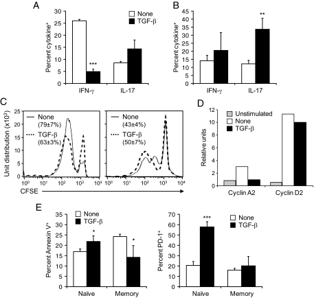
FIG. 5.
TGF-β suppresses naïve CD8+ T-cell activation and enhances the survival and effector function of antigen-experienced/memory CD8+ T-cells. CD8+ T-cells were purified from the spleen of naïve P14 or LCMV-immune C57BL/6 mice, labeled with CFSE, cultured with GP33-loaded dendritic cells in the presence or absence of 3 ng/ml rhTGF-β1, and harvested 2 days later. A and B: Cells were analyzed for IFN-γ and IL-17 production by flow cytometry after stimulation with GP33. Data represents the average frequency of naïve (A) and memory (B) CD8+ T-cells producing IFN-γ or IL-17 ± SD from three individual mice per group. C: Cell proliferation was assessed by measuring CFSE dilution by flow cytometry. Representative fluorescence-activated cell sorting plots are shown. Values show the percentage of naïve (left panel) or memory (right panel) CD8+ T-cells that have undergone at least one division on stimulation in the presence or absence of TGF-β ± SD from three individual mice per group. D: Naïve GP33-specific CD8+ T-cells were analyzed for cell cycle protein RNA by RNase protection assay as described in Fig. 2B. E: Cells were analyzed by flow cytometry for apoptosis after staining with annexin V or for expression of PD-1. Data represents the average frequency of CD8+ T-cells stained with annexin V (left panel) or expressing PD-1 (right panel) ± SD from three individual mice per group.

