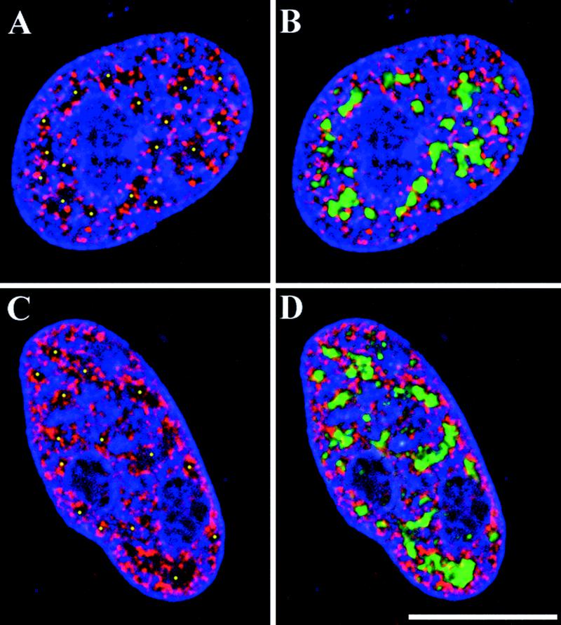
Figure 2.
Organization of dynamically acetylated euchromatin in the fibroblast nucleus. Digital optical sections of 0.4 μm were collected from Indian muntjac fibroblast cell nuclei that were cultured in the absence (A and B) or presence (C and D) of 10 mM sodium butyrate for 60 min. Cells were stained with an antibody specific to highly acetylated histone H3 (red), anti-SC-35 (green), and DAPI (red). The yellow dots in A and C indicate the positions of chromatin-depleted regions of the cell nucleus that correspond to regions positive for SC-35 (C and D). Bar, 10 μm.

