Abstract
Eighteen patients with proximal tibial shaft non-union and shortening were treated. In each patient, the non-union area was débrided, realigned and stabilised with an Ilizarov lengthening frame. The tibia was gradually lengthened by 1–1.5 mm per day. After achieving the desired length, external fixation was converted to an angled blade plate and packed with cancellous bone graft. Follow-up of 16 patients for a median of 2.4 (1.2–4.5) years revealed satisfactory outcomes in all. No wound infections were noted. The described technique has a high success rate, a short treatment course and reduces patient discomfort. This method may be considered preferential treatment for all patients with the specified indications.
Résumé
18 patients présentant une pseudarthrose de la diaphyse tibiale, avec raccourcissement ont été traités. Pour chaque patient, la pseudarthrose a été mise à plat avec correction d’axe et stabilisation avec un appareil d’Ilizarov. Le tibia a été allongé progressivement de 1 à 1.5 mm par jour. Après correction de l’inégalité de longueur, la fixation externe a été remplacée par une lame plaque associée à une greffe. 16 patients ont été traités ainsi avec un suivi moyen de 2.4 ans (1.2 à 4.5). Il n’y a pas eu d’infections profondes. Cette technique entraîne un taux de succès important avec un traitement plus rapide et une amélioration du confort des patients. Cette méthode peut être considérée comme adaptée à tous les patients.
Introduction
Proximal tibial shaft non-union with shortening is rare and normally caused by failed treatment of acute fractures. Although treatment options vary, no surgical technique is clearly superior. Each technique has individual advantages and disadvantages.
Tibial lengthening is generally favoured as a gradual technique using an external fixator (EF) to avoid neurovascular compromise [5, 17]. However, once the tibia achieves the desired length, several techniques may be employed to achieve bony union. The EF may be maintained in position, but an extended period may be required to achieve satisfactory results [3]. Thus, the patient may suffer more discomfort. Cancellous bone graft may speed the healing process, but the success rate may be compromised by insufficient stability of the EF [6, 7, 13]. Converting to internal fixation is suggested when necessary to reinforce local stability and increase the union rate [4, 6]. However, the nailing system is unsuitable in the proximal tibia due to the insufficient bony stock. Buttress plates may not be sufficiently stable due to the short length of proximal bony stock and local osteoporosis. The purpose of this retrospective study was to report the experience of this surgical team using an Ilizarov frame to enhance the speed of gradual lengthening and subsequently conversion to angled blade plate stabilisation.
Materials and methods
From May 1999 to December 2004, 18 consecutive adult (>15 years) patients with proximal tibial shaft non-union and shortening were treated by the described technique at our institution. A non-union was defined as a fracture remaining unhealed after 1 year of treatment or requiring additional surgery to achieve union [20]. Patients were aged a median of 32 years (19–54) with a male to female ratio of 5:1 (Table 1).
Table 1.
Clinical data for 18 patients with proximal tibial shaft non-union and shortening
| Case No. | Sex | Age (years) | Period since injury (years) | Operating times | Pre-op shortening (cm) | Pre-op PMTA (°) | Post-op PMTA (°) | Union period (months) | Last PMTA (°) | Functional | Follow-up (years) |
|---|---|---|---|---|---|---|---|---|---|---|---|
| 1 | M | 38 | 1.2 | 0 | 2.5 | 89 | 90 | 4.0 | 88 | G | 4.5 |
| 2 | M | 26 | 2.2 | 1 | 3.0 | 85 | 88 | L | L | L | L |
| 3 | M | 21 | 1.1 | 3 | 3.0 | 88 | 88 | 4.5 | 88 | G | 4.2 |
| 4 | M | 41 | 2.4 | 0 | 3.5 | 70 | 88 | 4.0 | 88 | E | 4.0 |
| 5 | F | 39 | 2.2 | 1 | 3.0 | 82 | 89 | 3.0 | 86 | G | 3.5 |
| 6 | F | 32 | 1.8 | 2 | 3.0 | 84 | 88 | 5.5 | 86 | E | 3.2 |
| 7 | M | 40 | 2.0 | 1 | 2.5 | 85 | 88 | 4.5 | 87 | E | 2.8 |
| 8 | M | 32 | 2.8 | 3 | 3.0 | 78 | 87 | 5.0 | 85 | G | 2.7 |
| 9 | M | 23 | 1.8 | 0 | 2.5 | 83 | 88 | 3.0 | 88 | G | 2.5 |
| 10 | M | 19 | 1.5 | 0 | 3.0 | 88 | 89 | 3.5 | 88 | E | 2.4 |
| 11 | F | 20 | 3.0 | 1 | 3.0 | 85 | 89 | 3.0 | 87 | G | 2.3 |
| 12 | M | 54 | 3.4 | 2 | 4.0 | 85 | 89 | 4.0 | 88 | E | 2.1 |
| 13 | M | 26 | 2.1 | 1 | 2.5 | 80 | 88 | 4.0 | 87 | G | 2.0 |
| 14 | M | 27 | 1.6 | 2 | 2.5 | 87 | 87 | 4.5 | 87 | G | 1.8 |
| 15 | M | 25 | 1.2 | 0 | 2.5 | 88 | 88 | L | L | L | L |
| 16 | M | 48 | 4.5 | 2 | 2.5 | 84 | 88 | 3.0 | 87 | E | 1.5 |
| 17 | M | 30 | 1.4 | 0 | 3.0 | 84 | 89 | 5.0 | 87 | G | 1.5 |
| 18 | M | 38 | 1.6 | 0 | 3.0 | 82 | 90 | 4.0 | 88 | G | 1.2 |
E excellent, F female, G good, L lost to follow-up, M male, N non-union, PMTA proximal medial tibial angle, Post-op post-operative, Pre-op pre-operative
All non-unions were due to failed treatment of acute fractures, and all fractures were initially sustained in motor vehicle accidents. Three fractures were initially type 2 open fractures and the other 15 were closed fractures [8]. Initial fracture treatment included conservative treatment by bone setting in nine cases, casting in five and buttress plating in four. Due to failure of initial treatment, 11 patients underwent surgery and other 7 patients did not. Although 11 patients underwent one to three operations, all fractures remained unhealed. The median time from initial injury to the described revision surgery was 1.8 years (1.1–4.5). No wound infection occurred. The tibial shortening was a median of 3.0 cm (2.5–4.0). The proximal medial tibial angle (PMTA) was a median of 85° (70–89) (Table 1). All patients had a full range of motion (ROM) in the knee. Although crutches were not used, no patients could tolerate walking for extended periods.
In the outpatient department (OPD), the wound healing process was carefully monitored. Non-unions with past histories of deep infection were excluded and the EF was not replaced. Indications for the described technique included proximal tibial shaft aseptic non-union (distal to the tibial tubercle), shortening exceeding 2.0 cm and unsuitable anatomy for inserting proximal locking screws.
On admission, white blood cell count, erythrocyte sedimentation rate and C-reactive protein were routinely checked. Non-unions with suspicious latent infection were excluded, and the EF was maintained until union was achieved.
Surgical technique
Under spinal anaesthesia, patients were positioned on the operating table in the supine position. A pneumatic tourniquet was routinely used.
A mid-third fibulotomy was performed first. The tibial non-union site was approached directly from the anterior route. The prior implants were removed, and the local area was débrided. All scar tissues were completely excised. After realigning the tibial axis, an Ilizarov lengthening frame (TraumaFix, San Francisco, CA, USA) was inserted. The wound was closed with absorbable sutures.
Post-operatively, patients were permitted to ambulate with protected weight bearing immediately and were advised to let the foot of the affected leg lightly touch the ground. The pin tract was disinfected with 70% alcohol once a day. Lengthening was introduced at 1–1.5 mm per day (in 4–6 sessions) when wound pain was tolerable. Exercises for knee and ankle ROM were encouraged. When the tibia reached a length comparable to that of the contralateral tibia, patients were re-admitted for further surgical treatment. The median interval between the two operations was 25 days (21–32). No pin tract infection was noted.
Under general anaesthesia with endotracheal intubation, patients were placed on the operating table in the lateral decubitus position. Massive corticocancellous bone graft was first procured from the posterior iliac crest. The wound was closed with absorbable sutures.
Patients were then placed in the supine position and the EF was removed. A pneumatic tourniquet was applied. A skin incision was made along the previous wound and extended upwards. The local area was examined carefully and scar tissues were removed. A spinal spreader was applied to maintain the maximal length of the tibia. An adequately sized blade plate angled 95° (Synthes, Bettlach, Switzerland) was selected. The blade was inserted in the direction of the fibular head and at least four cortical screws were required for the distal tibial fragment. With the knee maintained at 7° of valgus alignment, the angled blade plate was applied on the anteromedial aspect of the tibia. After insertion of all screws, the spinal spreader was removed. Knee alignment, tibial length and local stability were checked. The knee was manipulated to ensure full ROM. After the bone graft was packed in the space, the wound was closed with non-absorbable sutures. A closed drain was routinely inserted.
Post-operatively, patients were encouraged to ambulate with protected weight bearing and to perform knee ROM exercise. They were advised to let the foot of the affected leg lightly touch the ground. Patients were followed-up in the OPD at 4- to 6-week intervals. Clinical and radiographic fracture healing processes were recorded. After the nonunion had healed, patients were followed up in the OPD annually or as necessary.
A fracture union was defined as: clinically lack of pain or tenderness, ability to walk unaided and radiographically solid callus bridging both fragments [18].
To assess the outcome, the treatment rules were divided into bone and functional categories, according to the classification of the Association for the Study and Application of the Method of Ilizarov [14]. Four grades were classified and an excellent or good result was considered satisfactory.
Results
Sixteen patients were followed up for a median of 2.4 years (1.2– 4.5); two patients could not be contacted (Table 1).
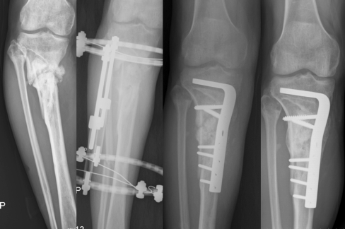
All non-unions healed within a median period of 4.0 months (3.0–5.5) after angled blade plate stabilisation (Fig. 1).
Fig. 1.
Case 4. A 41-year-old man sustained a right proximal tibial shaft non-union, varus deformity of 17° and shortening of 3.5 cm for 3.5 years. These combined lesions were treated with the described technique. The non-union healed at 4 months and an excellent result was achieved at the 4-year follow-up
No wound infection or malunion (shortening > 2 cm, angular or rotational deformity > 5°) was noted. Functional outcome revealed that before revision surgery all patients had unsatisfactory grades. However, at final follow-up, all patients had satisfactory grades. Concomitantly, all patients achieved normal gait and a full squat.
Knee alignment in all patients improved to within an acceptable range (PMTA: 80–94°) either immediately post-operatively or at final follow-up [16].
Implants were intended to be left in place unless the patient reported discomfort. No implants were removed as of the last follow-up.
Discussion
Factors favouring fracture healing include minimal gap, adequate stability and sufficient nutrition supply [11]. Once a non-union occurs, treatment principles include minimising the gap, providing stability and initiating osteogenic potential [15]. Although a non-union is traditionally classified as hypertrophic or atrophic, cancellous bone graft is recommended in both types of non-union to increase the union rate [12]. In the described technique, massive cortico-cancellous bone graft was supplemented and all non-unions healed.
Gradual lengthening is normally from a freshly osteotomised site. The general rule of lengthening is 1 mm per day [10, 14]. Sometimes, cancellous bone graft may be unnecessary. However, the possibility of spontaneous healing is minimal after lengthening from a non-union site. Therefore, cancellous bone grafting is essential. Now that bone graft must be supplemented, speeded gradual lengthening seems to be reasonable. Minimising the EF insertion period can reduce the discomfort caused by the procedure and decrease the complication rate of EF [6, 13]. In this study, the EF was placed with a median of 25 days (21–32), and no infection occurred. A literature review indicates pin tracts in EF are considered secure within 3 weeks [19].
Previous studies also indicate speeded gradual lengthening at 1.5 mm per day is considered secure and neurovascular injuries are not an issue [9, 17]. In this study, lengthening of 1–1.5 mm per day was achieved as long as the pain was tolerable. EF insertion periods are greatly reduced and thus pin tract infection is prevented.
The relatively short bone stock in the proximal fragment greatly restricts the use of locked nails. Osteoporosis near the non-union site severely endangers the stability of buttress plate screws. Angled blade plates have been successfully used to treat non-union of the tibial plateau [2, 16]. In this study, angled blade plates also proved useful for proximal tibial shaft lengthening.
Advantages of angled blade plates in treating proximal tibial shaft non-union and shortening are multiple. The blade has better stability to prevent implant migration in the osteoporotic bone [2, 16]. The blade also has a larger contact area with the bone to resist shortening. Compared to traditional non-locked plates, the fixed-angle blade plate is superior in maintaining the plate contour. The surgical technique is not complex. However, in a lengthening procedure, a large bony gap allows the plate to become a complete load-bearing device [1]. If patients do not ambulate with protected weight bearing, implants frequently fail. A co-operative patient is ultimately the key to successful treatment.
In conclusion, speeded gradual lengthening and secondary angled blade plate stabilisation is an excellent technique for treating proximal tibial shaft non-union with shortening. A high success rate can be confidently predicted in carefully selected patients. Additionally, the technique is not complex and employs commonly used devices. This method may be considered the preferred treatment for all patients with the specified indications.
References
- 1.Albright JA, Johnson TR, Saha S. Principles of internal fixation. In: Ghista DN, Roaf R, editors. Orthopedic mechanics: procedures and devices. London: Academic; 1978. pp. 123–229. [Google Scholar]
- 2.Carpenter CA, Jupiter JB. Blade plate reconstruction of metaphyseal nonunion of the tibia. Clin Orthop. 1996;332:23–28. doi: 10.1097/00003086-199611000-00005. [DOI] [PubMed] [Google Scholar]
- 3.Cierny G, III, Zorn KE. Segmental tibial defects: comparing conventional and Ilizarov methodologies. Clin Orthop. 1994;301:118–123. [PubMed] [Google Scholar]
- 4.Coleman SS, Stevens PM. Tibial lengthening. Clin Orthop. 1976;136:92–104. [PubMed] [Google Scholar]
- 5.Dahl MT, Gulli B, Berg T. Complications of limb lengthening: a learning curve. Clin Orthop. 1994;301:10–18. [PubMed] [Google Scholar]
- 6.Faber FWM, Keessen W, Roermund PM. Complications of leg lengthening: 46 procedures in 28 patients. Acta Orthop Scand. 1991;62:327–332. doi: 10.3109/17453679108994463. [DOI] [PubMed] [Google Scholar]
- 7.Green SA. Skeletal defects: a comparison of bone grafting and bone transport for segmental skeletal defects. Clin Orthop. 1994;301:111–117. [PubMed] [Google Scholar]
- 8.Gustilo RB, Anderson JTT. Prevention of infection in the treatment of one thousand and twenty-five open fractures of long bones: retrospective and prospective analysis. J Bone Joint Surg Am. 1976;58:453–458. [PubMed] [Google Scholar]
- 9.Hang YS, Shih JS. Tibial lengthening: a preliminary report. Clin Orthop. 1977;125:94–99. [PubMed] [Google Scholar]
- 10.Ilizarov GA. The tension-stress effect on the genesis and growth of tissues: part II. The influence of the rate and frequency of distraction. Clin Orthop. 1989;239:263–285. [PubMed] [Google Scholar]
- 11.Karlstrom G, Olerud S. Fractures of the tibial shaft: a critical evaluation of treatment alternatives. Clin Orthop. 1974;105:82–111. [PubMed] [Google Scholar]
- 12.LaVelle DG. Delayed union and nonunion of fractures. In: Canale ST, editor. Campbell’s operative orthopedics. St. Louis: Mosby; 2003. pp. 3125–3165. [Google Scholar]
- 13.Paley D. Problems, obstacles, and complications of limb lengthening by the Ilizarov technique. Clin Orthop. 1990;250:81–104. [PubMed] [Google Scholar]
- 14.Song HR, Cho SH, Koo KH, Jeong ST, Park YJ, Ko JH. Tibial bone defects treated by internal bone transport using the Ilizarov method. Int Orthop. 1998;22:293–297. doi: 10.1007/s002640050263. [DOI] [PMC free article] [PubMed] [Google Scholar]
- 15.Weber BG, Brunner C. The treatment of non-union without electrical stimulation. Clin Orthop. 1981;161:24–32. [PubMed] [Google Scholar]
- 16.Wu CC. Salvage of proximal tibial malunion or nonunion with the use of angled blade plate. Arch Orthop Trauma Surg. 2006;126:82–87. doi: 10.1007/s00402-006-0106-9. [DOI] [PubMed] [Google Scholar]
- 17.Wu CC, Chen WJ. Tibial lengthening: technique for speedy lengthening by external fixation and secondary internal fixation. J Trauma. 2003;54:1159–1165. doi: 10.1097/01.TA.0000046254.92637.19. [DOI] [PubMed] [Google Scholar]
- 18.Wu CC, Chen WJ, Shih CH. Tibial shaft malunion treated with reamed intramedullary nailing: a revised technique. Arch Orthop Trauma Surg. 2000;120:152–156. doi: 10.1007/s004020050033. [DOI] [PubMed] [Google Scholar]
- 19.Wu CC, Shih CH. Complicated open fractures of the distal tibia treated by secondary interlocking nailing. J Trauma. 1993;34:792–796. doi: 10.1097/00005373-199306000-00007. [DOI] [PubMed] [Google Scholar]
- 20.Wu CC, Shih CH, Chen WJ, Tai CL. High success rate with exchange nailing to treat a tibial shaft aseptic nonunion. J Orthop Trauma. 1999;13:33–38. doi: 10.1097/00005131-199901000-00008. [DOI] [PubMed] [Google Scholar]


