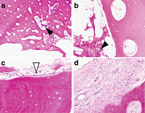
Fig. 5.
Histology of bone at prosthetic interface from three patients (a, b patient 8, c patient 2, d patient 3). Viable osteocytes are diffusely present in all three patients immediately adjacent to the medullary cavity. Viable bone marrow (solid arrowheads) and osteoblastic rimming (open arrowhead) are demonstrated around intraosseous vessels. Woven bone and loose fibrosis was also identified at the interface (d). Haematoxylin and eosin; original magnification 40× (a) and 200× (b–d)

