
Fig. 2.
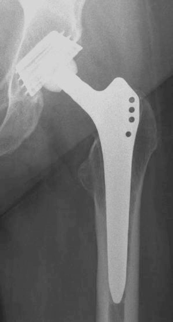
AP radiograph of the left hip of a 78-year-old female patient with rheumatoid arthritis 11 years after implantation of the Zweymueller prosthesis. Typical radiographic result with bone atrophy in the acetabulum in zones I and II, thin radiolucent lines around the stem in zones I and VII, and bone atrophy in the proximal femur in zones I and VII