
Fig. 6.
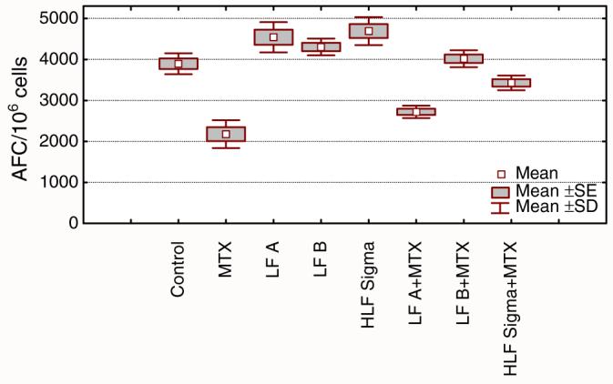
Reconstituting effect of lactoferrin on the secondary humoral immune response in mice suppressed by methotrexate. Splenocytes isolated from sheep red blood cell (SRBC) primed mice were incubated with SRBC, alone in the presence of methotrexate (MTX). Antibody forming cells (AFC) were evaluated after 4 days. Cells were cultured in the presence of non-sialylated LF (LF A), sialylated LF (LF B), or milk-derived LF (HLF Sigma). All LF concentrations were 1 μg/ml. The results are shown as mean values of AFC number from five wells±SE, calculated per 106 viable cells. Statistical analysis of groups: control vs. LF A NS; control vs. LF B NS; control vs. HLF Sigma p=0.0068; control vs. MTX p=0.0001; control vs. LF A+MTX p=0.0001; control vs. LF B+MTX NS; control vs. HLF Sigma+MTX NS; MTX vs. LF A+MTX NS; MTX vs. LF B+MTX p=0.0001; MTX vs. HLF Sigma+MTX p=0.0001 (ANOVA)