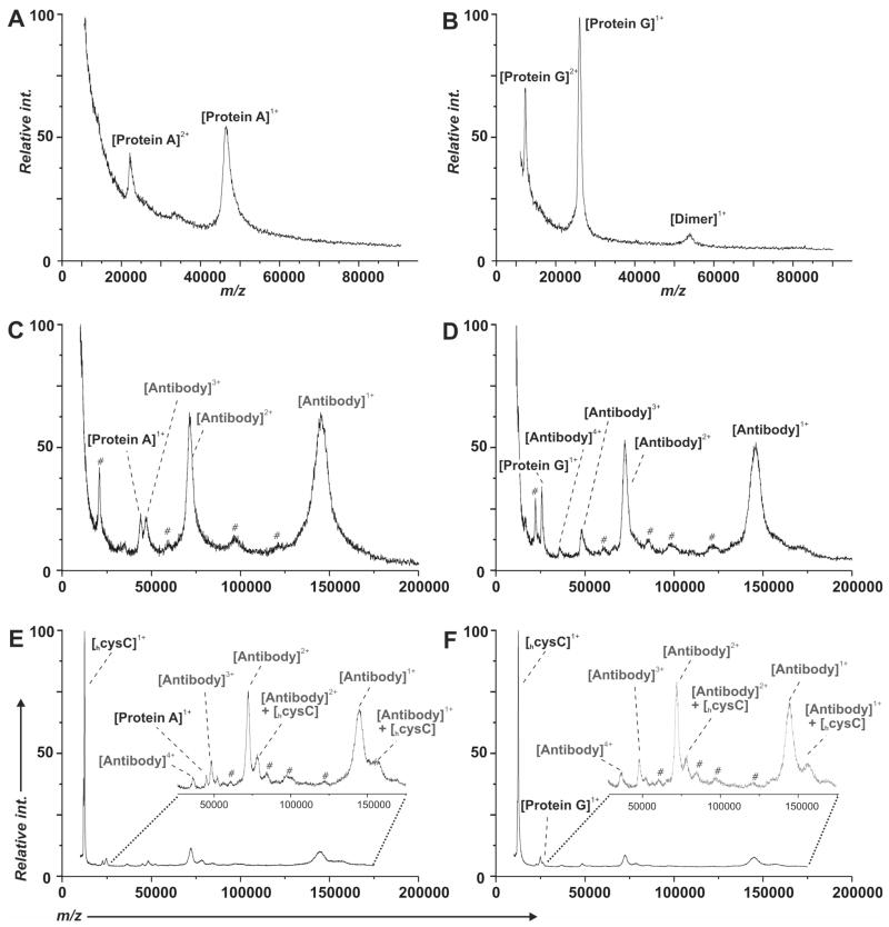
Figure 2.
SAMDI-TOF MS was used to monitor each step in the preparation of the immunosensor. The spectra in panels A and B show the immobilization of the protein A and protein G, respectively, onto a monolayer presenting the aza/Ni2+ ligand. Panels C and D show the immobilization of cysC antibody onto the protein A and protein G derivatized surfaces, respectively. Panels E and F show the capture of the cysC on the resulting immunosensors. (#) Unidentified components derived from the stock antibody.

