Figure 6.
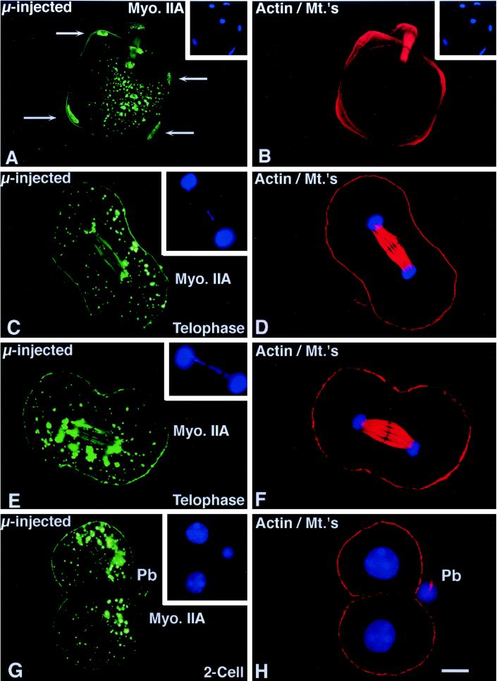
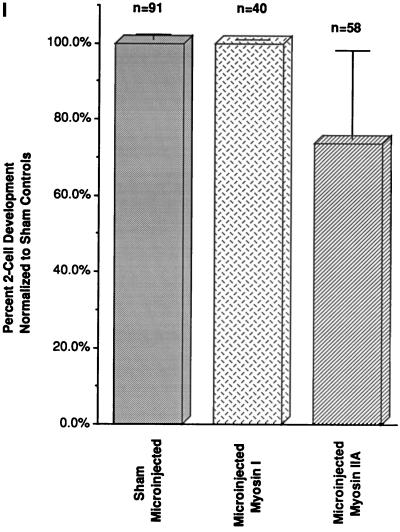
Effects of microinjected myosin IIA antibody on first mitosis. (A and B) Sperm incorporation in zona-free in vitro fertilized mature oocytes is not blocked in the presence of microinjected myosin IIA antibody. The assembly of cortical myosin IIA at the sperm incorporation cones appears reduced (A, arrows; compare with Figure 5A). Immunoreactive cytoplasmic aggregates are observed and both the paternal (A, arrows) as well as maternal chromatin bind the microinjected myosin IIA antibody. (C–F) Microinjection of myosin IIA into interphase zygotes does not impair chromosome congression, segregation, and cleavage furrow formation during mitosis. Cortical myosin IIA is not uniform and the intensity of antibody staining is reduced, especially within the forming cleavage furrow. Numerous cytoplasmic aggregates are observed and the labeling of mitotic chromosome is also evident. An occasional lagging chromosome is observed during late anaphase within the midbody region (insets). (D and F) Simultaneous antiactin and acetylated α-tubulin labeling demonstrates midbody and cortical microfilaments labeling in the same zygotes. (G and H) Cleavage of zygotes microinjected with myosin IIA appears to be normal and at the correct time for division. Detection of cortical myosin IIA in sister blastomeres is reduced in the cell–cell contact regions. No myosin IIA is found within daughter cell nuclei after cytokinesis, suggesting a transient association of myosin IIA with the condensed chromosomal surfaces during mitosis. H is the corresponding antiactin and acetylated α-tubulin image of the oocyte in G. (I) To quantify the effects of myosin II antibodies on cell division, pronucleate stage oocytes were microinjected with myosin I (I, middle column) or myosin IIA antibody (I, right column) and allowed to develop in vitro. Neither myosin antibody significantly impacts cytokinesis and two-cell formation following mitosis. Similar observations were made following microinjection of myosin IIB antibody, either injected alone or coinjection with the IIA antibody (our unpublished results). Left column, sham controls. Confocal images quadruple labeled for microinjected myosin IIA (green), actin, and acetylated α-tubulin (red) and DNA (blue). Bar, 10 μm.


