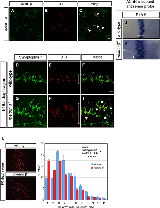
Figure 1. Meltrin β is expressed at the NMJ and participates in the formation of the NMJ.
(A–C) Adult tibialis anterior (T.A.) muscles were stained with an anti-Meltrin β antibody (A) and Alexa-594–conjugated α-bungarotoxin (BTX) (B). Meltrin β was expressed at NMJs (C, arrowheads). Meltrin β was also expressed in a part of muscle fibers. Bar: 10 µm. (D–I) The diaphragms of wild-type (D–F) and meltrin β −/− (G–I) mice at E18.5 were stained with an anti-synaptophysin antibody (D and G) to label axon terminals and with BTX (E and H) to label acetylcholine receptors (AChRs). AChR clusters were distributed more broadly (H and I, also compare the bars in E and H) and the nerve terminals sprouted excessively (G and I, arrowheads) in the meltrin β −/− muscle compared with in the wild-type muscle (F). Bar: 100 µm. (J and K) In situ hybridization with probes for AChR α-subunit mRNA in E18.5 diaphragms of wild-type (J) and meltrin β −/− (K) mice. The central zone expressing the AChR α-subunit mRNA is broader in meltrin β −/− (K) than in the wild-type diaphragm (J) (compare the bars in J and K). (L) Neonatal (P0) diaphragms were stained with BTX. AChR clusters were distributed more broadly in meltrin β−/− than in wild-type diaphragms (left panel). The average size of the individual AChR clusters was smaller in meltrin β−/− muscles relative to that in wild-type muscles (right panel). Bar: 100 µm.

