Figure 3.
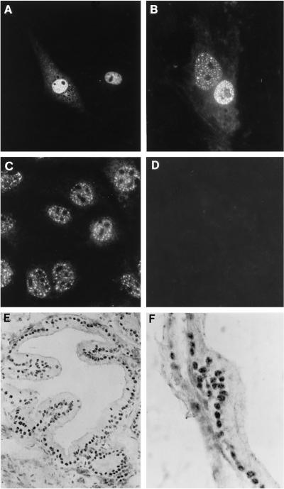
Localization of ANPK in cultured cells and prostate. (A and B) CV-1 cells seeded on glass coverslips on 10-cm plastic plates were transfected with 1 μg of FLAG-ANPK(2–1191) expression vector using DOTAP transfection reagent as described in MATERIALS AND METHODS. Cells were fixed in 4% (wt/vol) paraformaldehyde, permeabilized, and ANPK antigen was visualized using (A) affinity-purified rabbit antiserum raised against GST-ANPK(766–920) or (B) anti-FLAG M2 antibody. (C) The distribution of endogenous ANPK in S115 cells as determined by using anti-ANPK antiserum. (D) Immunofluorescence of S115 cells with anti-ANPK antiserum neutralized with purified GST-ANPK(766–920) fusion protein. (E and F) Distribution of ANPK in rat prostate. Immunoperoxidase technique was applied to visualize ANPK antigen using rabbit antiserum raised against GST-ANPK(766–920). (E) ANPK is localized in nuclei of epithelial cells. (F) A higher magnification showing granular clusters of ANPK immunoreactivity in epithelial cell nuclei. Original magnification; E, ×760; F, ×1,900.

