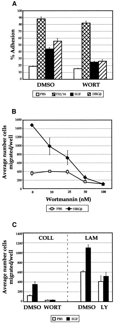
Figure 10.
PI 3-K inhibitors block EGF- or HRGβ-mediated adhesion and migration of MDA-MB-435 cells. (A) Adhesion assays were carried out in the presence of 100 nM wortmannin (WORT) with no stimulation (open bars) or after stimulation for 10 min at 37°C with TS2/16 (cross-hatched bars), EGF (shaded bars), or HRGβ(hatched bars). (B) Migration analysis was carried out in the presence of increasing amounts of wortmannin in the presence of media only (open squares) or HRGβ (solid triangles). Similar effects were observed when dose–response analysis was carried out on COLL or when assays were performed in the presence of 25 μM LY294002 (our unpublished data). (C) Migration on COLL or LAM was performed in the presence of control DMSO, 100 nM wortmannin (WORT), or 25 μM LY294002 (LY) with no stimulation (open bars) or stimulation with 10 ng/ml EGF (solid bars). HRGβ stimulation was slightly lower than normal in the adhesion assay shown in A in comparison with EGF or TS2/16 stimulation, but the data shown are otherwise representative of at least three separate experiments.

