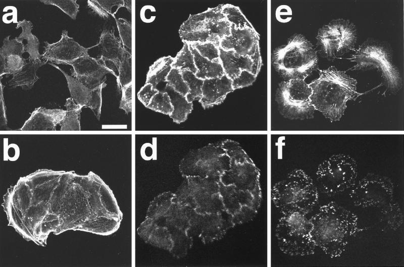
Figure 2.
Effect of cycloheximide on the TPA- and HGF/SF-induced reorganization of actin filaments in wt MDCK cells. wt MDCK cells were incubated in DMEM containing 10% FCS for 24 h. After the incubation, the cells were stimulated with 20 ng/ml HGF/SF (a and b) or 100 nM TPA (c–f) in the absence (a) or presence (b–f) of 10 μg/ml cycloheximide. Cycloheximide was added 30 min before HGF/SF or TPA stimulation. At 18 h after HGF/SF stimulation, the cells were fixed and stained with rhodamine-phalloidin (a and b). At 15 min (c and d) or 2 h (e and f) after TPA stimulation, the cells were fixed, double stained with rhodamine-phalloidin (c and e) or the V115 anti-vinculin mAb (d and f), and analyzed by confocal microscopy. Confocal images are shown at the basal levels. The results shown are representative of three independent experiments. Bar, 10 μm.

