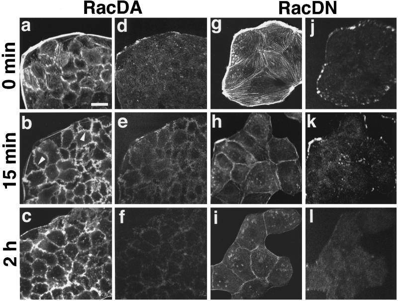
Figure 5.
Inhibition of the TPA-induced reassembly of stress fibers and focal adhesions in sMDCK-RacDA and -RacDN cells. sMDCK-RacDA and -RacDN cells were incubated in DMEM containing 10% FCS for 24 h. After the incubation, sMDCK-RacDA (a–f) and -RacDN (g–l) cells were stimulated with none (a, d, g, and j) or 100 nM TPA (b, c, e, f, h, i, k, and l). At 15 min (b, e, h, and k) or 2 h (c, f, i, and l) after TPA stimulation, the cells were fixed, double stained with rhodamine-phalloidin (a–c and g–i) or the V115 anti-vinculin mAb (d–f and j–l), and analyzed by confocal microscopy. Confocal images are shown at the basal levels. The results shown are representative of three independent experiments. Bar, 10 μm. Arrowheads in panel b indicate membrane ruffling at the cell–cell adhesion sites.

