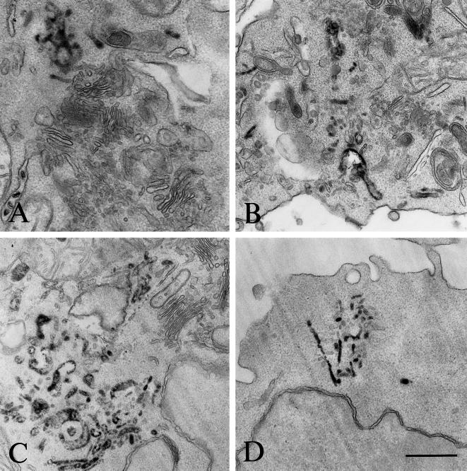
Figure 3.
Morphology of the HRP-containing compartments. Cells that had internalized HRP-Tf for 2 h at 19°C were processed for HRP detection using DAB and H2O2 and embedded in Epon. Ultrathin sections were then analyzed by electron microscopy. (A and B) B4.14 cells. (C and D) Laz cells. DAB reaction precipitates are observed in tubulovesicular structures. Bar, 500 nm.

