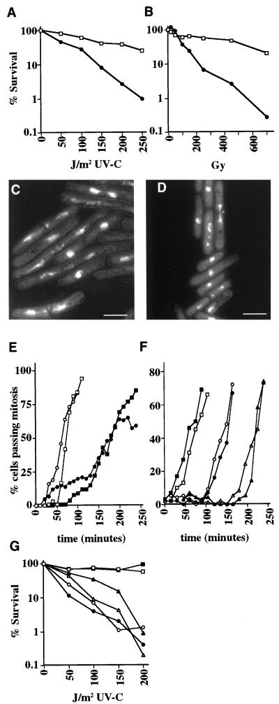
Figure 1.
NA74 is defective in the response to DNA damage. (A) UV-C survival curve for wild type (□) and NA74 (●). (B) Ionizing radiation survival curve for wild type (□) and NA74 (●). (C) DAPI-stained cells for NA74 that have been synchronized by cdc25-22 block and release and irradiated with 50 J/m2 UV-C. Bar, 10 μm. (D) DAPI-stained cells for asynchronous NA74 irradiated with 450 Gy ionizing radiation. Bar, 10 μm. (E) Wild-type cells (▪ and □) and NA74 (● and ○) were synchronized by cdc25-22 block and release and then irradiated with 50 J/m2 UV-C (closed symbols) or mock irradiated (open symbols). Cultures were incubated at 25°C, and ethanol-fixed samples were taken for DAPI staining. The percentages of cells that had passed mitosis were assessed from each sampleby fluorescence microscopy. (F) Wild-type cells (closed symbols) and NA74 (open symbols) were synchronized by lactose gradient centrifugation and then irradiated with 100 Gy (● and ○) or 500 Gy (▴ and ▵) or mock irradiated (▪ and □). Cultures were incubated at 30°C, and ethanol-fixed samples were taken for DAPI staining. The percentages of cells that had passed mitosis were assessed from each sample by fluorescence microscopy. (G) cdc25-22 (▪ and □), cdc25-22 NA74 (● and ○), and cdc25-22 rad3-136 (▴ and ▵) were grown at 25°C, plated on YES medium, and irradiated with the indicated dose of UV-C. Plates were then placed at 25°C (open symbols) or at 36°C for 4 h and then shifted to 25°C (closed symbols). Surviving colonies, expressed as a percentage of unirradiated controls, were counted after 4 d.

