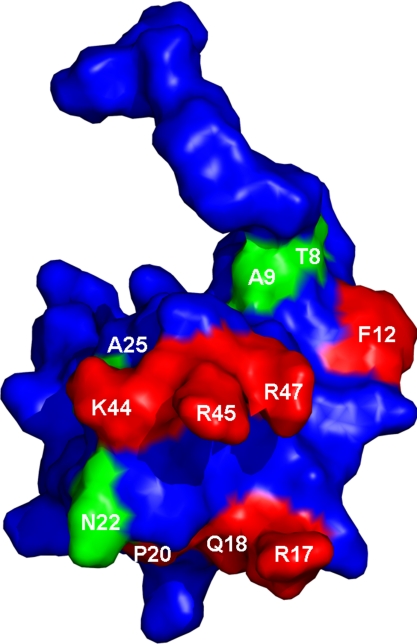
Fig. 5.
Three-dimensional structure of MIP-1α. Spatial orientation of residue side chains as derived from Czaplewski et al. (4), showing in red side chain residues that have been reported previously to be involved in CCR5 binding to structurally related MIP-1β (F12, R17, Q18, P20, K44, R45, and R47). Interactions identified from this study are highlighted in green (T8, A9, N22, and A25).

