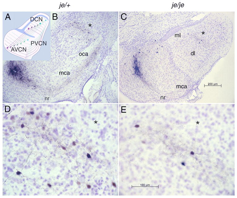
Figure 1.
Tonotopic organization of the cochlear nuclei is unaltered by the jerker mutation. A: A schematic representation of the topographic organization of the tuberculoventral cell projections from the dorsal (DCN) to the anteroventral (AVCN) and posteroventral cochlear nucleus (PVCN) that underlies the labeling pattern. A sheet of granule cells separates the ventral from the dorsal cochlear nucleus (blue); granule and other cell bodies separate the outer, molecular layer from the innermost deep layer (blue). Auditory nerve fibers impose a tonotopic organization on both the VCN and DCN; those fibers that encode the lowest frequencies (brown) terminate ventrally and those that encode the highest frequencies (blue-green) terminate dorsally. Tuberculoventral cells project to targets in the AVCN and PVCN that receive input from the same group of auditory nerve fibers and are therefore tuned to similar frequencies. (The topographic projection of T stellate cells to the DCN is not illustrated.) B: Photomicrograph of a parasagittal section of a slice of the cochlear nuclei from a heterozygous, je/+, normally hearing mouse in which an injection of biocytin had been made into the AVCN. The injection labeled a cluster of auditory nerve fibers that can be traced retrogradely from the injection site to the nerve root (nr) and anterogradely through the multipolar (mca) and octopus cell areas (oca) in the PVCN into the deep layer (dl), but not the molecular layer (ml), of the DCN. C: In a homozygous, deaf, je/je mouse the labeling pattern resembles that of the heterozygote. The oca is not present in this lateral section. D, E: Labeled fibers and cell bodies in the deep layer of the DCN in the same sections illustrated in B and C are shown at higher magnification, with asterisks (*) indicating corresponding points in panels B and D and in panels C and E. Within the bands of labeled fibers in the deep layer of the dorsal cochlear nucleus (DCN) lay bands of tuberculoventral cell bodies that were labeled through their terminals at the injection site. A few labeled cell bodies ventral to the band of labeled fibers were labeled through axons that passed through the injection site en route to more ventral regions of the AVCN.

