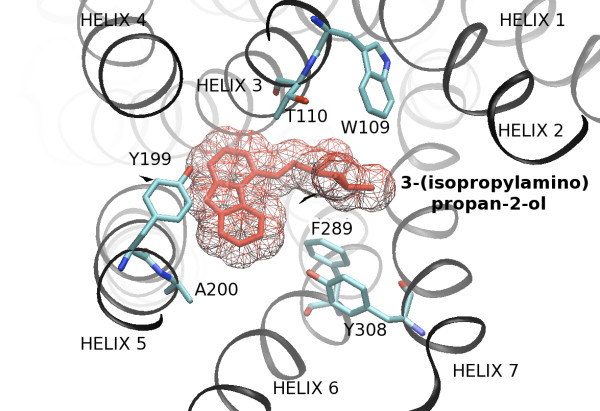
Figure 2.
3-(isopropylamino)propan-2-ol and the protein environment of β2-adrenergic receptor as viewed from the extracellular surface. 3-(isopropylamino)propan-2-ol and the protein environment of β2-adrenergic receptor as viewed from the extracellular surface. Amino acid side chains are represented for 6 of the 31 residues (in cyan, blue and red) of the binding pocket motif. Transmembrane helix and 3-(isopropylamino)propan-2-ol are colored in black and red respectively. Figure drawn with VMD [79].

