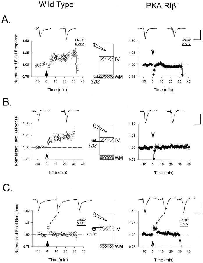
Fig. 3.
Defective LTP of extracellular field responses in the visual cortex of PKA RIβ− mice. TBS (arrow) applied (A) to the white matter (n = 6 and 5 slices from 4 and 3 mice, WT and RIβ−, respectively) or (B) directly to layer IV (n = 8 and 11 from 7 and 6 mice, WT and RIβ−, respectively) potentiates supragranular field response amplitudes in WT (○) but not RIβ− (•) mice recorded blind to genotype.C, More powerful tetani (four 1 sec bursts of 100 Hz;arrow) fail to induce LTP in both WT and mutant slices (n = 5 slices from 3 mice each). Representative traces 5 min before and 25 min after conditioning stimuli are shownabove each graph. Sample traces during post-tetanic potentiation are also indicated in C. Except for the experiments in B, which were continued to examine depotentiation (compare Fig. 6A), glutamate receptor antagonists CNQX (10 μm) and D-APV (50 μm) were routinely bath-applied to determine the synaptic nature of the field response. Calibration: 0.3 mV, 20 msec for each.

