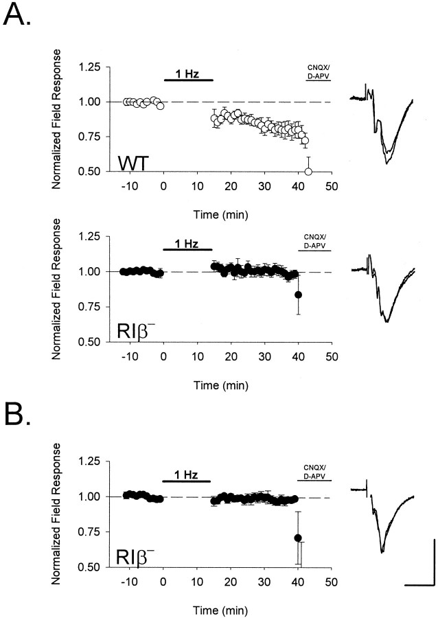
Fig. 6.
Absence of synaptic depression after low-frequency stimulation in PKA RIβ− mice. Extracellular field potential amplitude was monitored in layer II/III after low-frequency stimulation (900 pulses at 1 Hz) to layer IV of visual cortex (•, RIβ−; ○, WT). A, Renormalized responses after an earlier TBS (compare Fig. 3B) were depotentiated in wild-type (n = 8 slices from 7 mice) but unchanged in RIβ− mice (n= 11 slices from 6 animals). B, Low-frequency stimulation was similarly ineffective at naïve RIβ− synapses (n = 5 slices from 3 mice). Bath application of CNQX (10 μm) and D-APV (50 μm) terminated each experiment to confirm the synaptic nature of the field response. Representative traces 5 min before and 20 min after LFS are shown superimposed to the right of each graph. Calibration: 0.3 mV, 10 msec.

