Fig. 3.

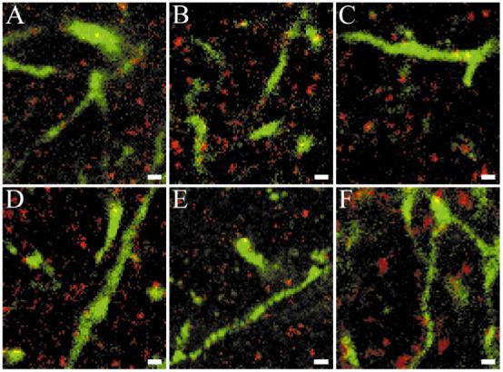
Visual image inspection is inadequate to determine whether TrkB-like immunoreactivity is colocalized with geniculocortical afferents. False-color double-label immunofluorescent confocal images from layer IV of kitten visual cortex are shown in red for TrkB23 (A), TrkB146 (B), TrkB348 (C), TrkB606 (D), RTB (E), and GAD65 (F) and in green for Phaseolus vulgaris leucoagglutinin (PHA-L)/-labeled geniculocortical afferent arbors (A-F). Although apparent colocalization of TrkB-positive puncta with PHA-L-labeled afferents can be observed for all antibodies, it is also present for GAD65. Scale bars = 1 μm in A-F.
