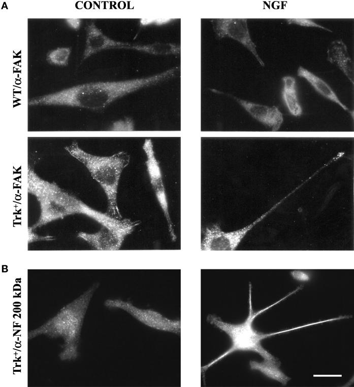
Figure 4.
(A) Immunofluorescence analysis of FAK expression. Wild-type (WT) or trk-PC12 (Trk+) cells fixed under control conditions (left panels) or after treatment with NGF for 24 h (right panels) were analyzed for expression of focal adhesion kinase (α-FAK). Increased levels of FAK immunostaining can be observed in trk-PC12 cells, partially colocalizing with structures similar to focal adhesions under control conditions and relocalizing to the growth cone after 24 h of NGF treatment. (B) Immunofluorescence analysis of neurofilament expression and localization in trk-PC12 cells. Control and NGF-treated trk-PC12 cells were analyzed for expression of the 200-kDa neurofilament subunit (α-NF 200 kDa). Although under control conditions the protein is expressed at low levels and appears diffuse (left panel), it is strongly up-regulated after NGF treatment and enriched in the growing processes (right panel). Bar, 14 μm.

